Craniotomy Posterior Fossa. Discrete posterior fossa cerebrospinal fluid CSF collections that are clearly separate from the fourth ventricle and vallecula are classified as posterior fossa arachnoid cysts PFAC12 Arai and Sato had clearly outlined the indications for surgery in PFAC.
Posterior Cranial Fossa Boundaries Contents Teachmeanatomy
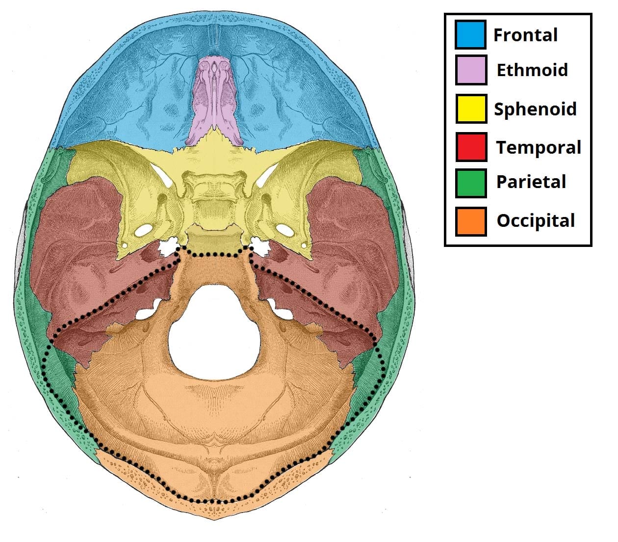
Bones forming the posterior cranial fossa by Anatomy Next.
. Gastrointestinal stromal tumor very rare 5. The posterior cranial fossa is comprised of three bones. Posterior middle and anterior.
Surgery in the areas to the rear of the skull also known as the posterior fossa or cerebellum can cause long lasting negative effects in children. It is bounded as follows. Cerebellar metastases most common.
With half being found in the posterior fossa Cerebellar astrocytomas make up 40 of pilocytic atrocytomas Usually occur in the latter half of the first decade Mean age of 7 years old Rarely found in children less than 1 year of age. A postoperative syndrome labeled posterior fossa syndrome has been identified in certain children. These areas of the brain control the autonomic nervous system coordination.
The posterior fossa is a small space in the skull found near the brainstem and cerebellum. Borders of posterior cranial fossa. Most common posterior fossa primary brain tumor in adults.
Aneurysm AVM SAH Tumor Chiari Pre-op Neurologic condition. Posterior fossa meningiomas are tumors that form near the bottom of the skull by the brainstem and cerebellum. Is a condition that sometimes develops after surgery to remove a brain tumor in the posterior fossa region of the brain.
This is a large superior projection of bone that arises from the body of the sphenoid. Posterior fossa meningiomas that impinge on structures of the temporal bone or clivus may be difficult to access for optimal resection that maximizes tumor control and minimizes short- and long-term morbidities. 11cThe trigeminal nerve exits the posterior fossa through an ostium formed by a.
The posterior fossa accommodates the cerebellum and brain stem. These effects include loss of muscle tone memory troubles unsteadiness and decreased ability to talk. Management strategy depends upon the size and location of these arachnoid cysts within the posterior fossa.
The posterior fossa or posterior cranial fossa is the deepest and largest and is defined by the occipital bone of the skull. Within this fossa are two critical brain areas. The brainstem is responsible for controlling vital body functions such as breathing.
Fossa cranii posterior lies at the lowest level of the internal cranial base and is the largest of the three cranial fossae. The bones of the posterior fossa are lined with dura mater which often contains venous lakes between its layers. The posterior fossa is a small space in the skull found near the brainstem and cerebellum.
The brainstem is responsible for controlling vital body functions such as breathing. Posterior fossa syndrome PFS or cerebellar mutism syndrome CMS is a collection of neurological symptoms that occur following surgical resection of a posterior fossa tumour and is characterised by either a reduction or an absence of speech. Preoperative Evaluation and Questions.
This small area controls movement coordination and. If a tumor grows in the area of the posterior fossa it can block the flow of spinal fluid and cause increased. To address this challenge the contemporary neurosurgeryneurotology team works collaboratively by managing patients jointly at every stage of care.
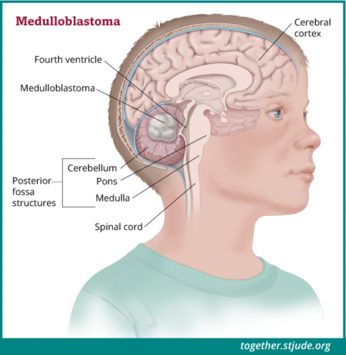
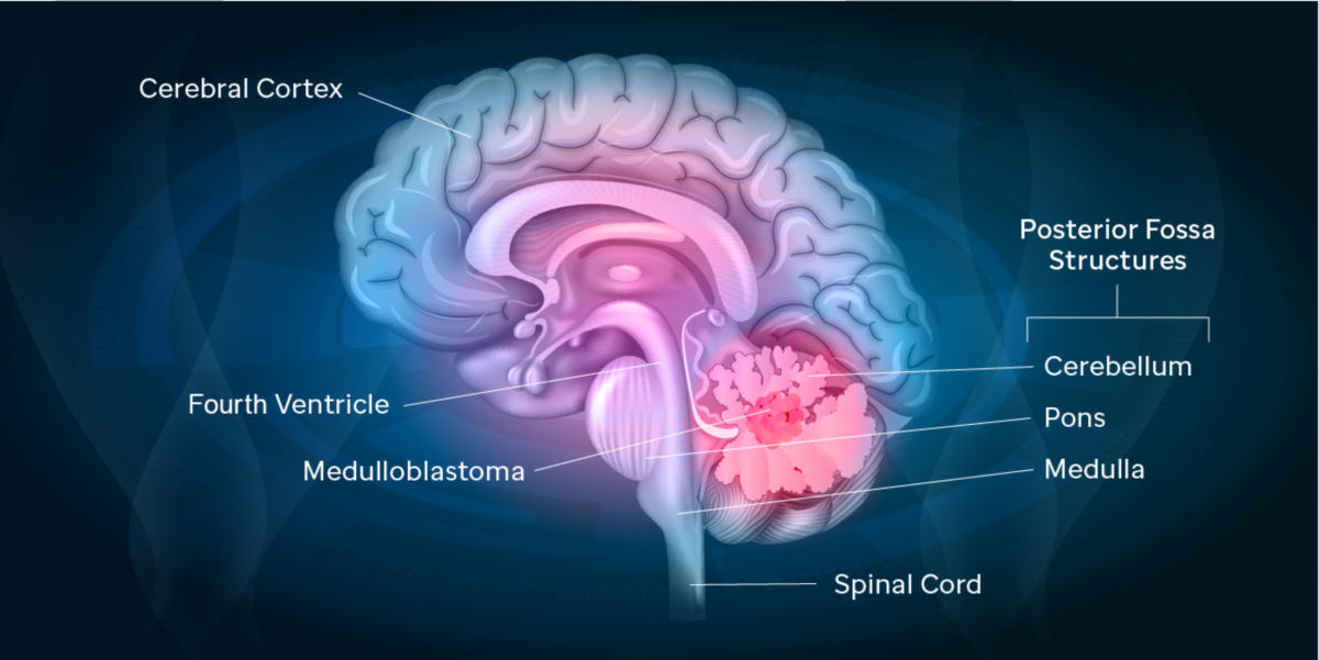
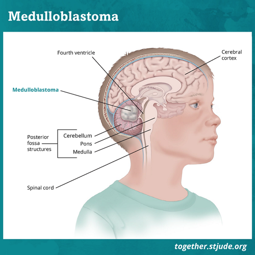
PFA or midline floor of the fourth ventricle near the obex molecular subgroup. Its is formed by the sphenoid temporal and occipital bones. Medulloblastoma the most common malignant brain tumor of childhood occurs in the posterior fossa the part of the intracranial cavity that contains the brainstem and the cerebellum.
Herewith the authors present a patient with a ruptured aneurysm of the anterior spinal artery associated with dural AVM of the posterior fossa. The occipital bone and the two temporal bones. Superiorly the cerebellum is separated from the cerebral hemispheres by the tentorium cerebelli.
The cerebellum is involved in many complex aspects of human behavior and function and when it is disrupted or insulted this can lead to significant sequelae in children with posterior fossa. Approximately 60 to 70 of pediatric brain tumors originate in the posterior fossa. The base of the skull is divided into three cranial fossae.
Hunt-Hess Grade for Aneurysm. Posterior fossa ependymomas are apt to extend through the foramina of Luschka and Magendie hence the term plastic ependymoma. The majority of posterior fossa ependymomas arise from the lateral recess of the fourth ventricle molecular subgroup.
Posterior fossa syndrome typically occurs in children but early detection of the condition and ensuing treatment may help reduce the need for lengthy hospital stays. The brain stem and the cerebellum. Also melanoma thyroid malignancies and renal cell cancer.
These effects are collectively known as Cerebellar Mutism Syndrome CMS or Posterior Fossa Syndrome. The Posterior Fossa Society is an international group of researchers and health care professionals doctors nurses psychologists speech pathologists linguists neuroscientists etc who are dedicated to research into the causes features treatment and prevention of the post-operative pediatric CMS and CCAS in children and adults. Anteriorly and medially it is bounded by the dorsum sellae of the sphenoid bone.
Since 1989 the two hospitals that comprise the setting for this study have treated 121 children with posterior fossa brain tumors. What is the indication for surgery. The posterior cranial fossa Latin.
7 Symptoms typically begin with intermittent headache often worse in the morning followed by vomiting and eventually gait disturbance. The posterior fossa is a space near the base of the skull that contains the. The posterior fossa region contains the brainstem which is responsible for controlling breathing regulating heart rate dilating and constricting blood vessels and giving a person the ability to.
The incidence of an aneurysm of the spinal artery is extremely rare. Anteriorly and laterally there are five pairs of neural foramina into which a sleeve of posterior fossa dura follows forming small or large CSF-filled dural caves Fig. The cerebellum is the part of the brain responsible for balance and coordinated movements.
Posterior fossa tumor has a very different differential in an adult as opposed to a child. Only several cases have been documented previously. If a tumor grows in the area of the posterior fossa it can block the flow of spinal fluid and.
Some authors suggest that CM is only one symptom of the CMS complex that also includes ataxia. Especially lung cancer and breast cancer. Posterior fossa tumors often cause hydrocephalus by blocking cerebrospinal CSF outflow pathways with resulting signs and symptoms of raised intracranial pressure ICP.
The cerebellum is the part of the brain responsible for balance and coordinated movements. Posterior fossa - axial CT image at level of pons. Children with posterior fossa syndrome usually have a collection of symptoms.
Getting To The Bottom Of A Medulloblastoma Mystery St Jude Progress
Posterior Fossa Syndrome Together
Posterior Fossa Syndrome Together
Epos C 0323

Posterior Cranial Fossa Wikipedia

Posterior Fossa Anatomy Operative Neurosurgery

Anatomical Location Of Posterior Fossa Meningiomas Cerebellar Download Scientific Diagram

Posterior Cranial Fossa Radiology Reference Article Radiopaedia Org
0 comments
Post a Comment